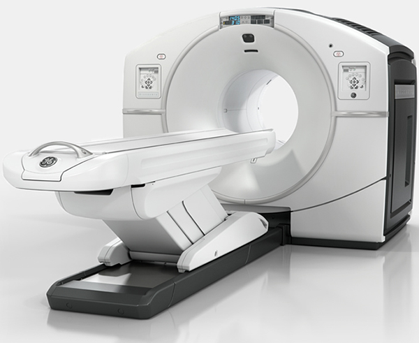
GE/Discovery IQ PET CT – First in South Tamilnadu to have a PET CT
PET CT is revolutionizing Cancer care
The value of Whole body Positron Emission Tomography–Computed Tomography (PET/CT) for diagnostic imaging in oncology is well established with its capabilities to combine anatomical and functional aspects of the whole body to provide a complete picture of patient’s disease status.
PET CT can help speed up treatment, because metabolic changes in a tumor occur more frequently than structural changes. It is a more accurate and reliable solution that can help clinicians determine how well a treatment is working especially in lymphomas after as few as 1 to 2 cycles of chemotherapy, and they will be able to tailor the treatment according to individual patient’s response and needs. Thus Molecular imaging using PET CT is a decisive tool for early diagnosis, treatment staging as well as monitoring the progress of treatment, while also reducing overall treatment cost.
Most common uses of PET/CT are found in Oncology, Cardiology and Neurology practices.
- FDG PET/CT is an integral part of cancer management in Evidence Based Practice. PETCT is used for initial staging of various cancers, Radiotherapy planning, treatment response assessment, restaging and surveillance in Oncology practice.It changes treatment decision in significant proportion of cancer patients.
- In Cardiology practice, FDG PET/CT is the gold standard test for detection of myocardial viability. It provides evidence for selection of heart disease (coronary artery disease) patients for revascularization (stenting / bypass surgery).
- In Neurology practice, FDG PET/CT is used to provide differential diagnosis for Dementia it helps Neurosurgeons in mapping the brain for surgical resection, in cases of drug resistant Epilepsy.
Linac with IGRT, IMRT, Rapid arc facility
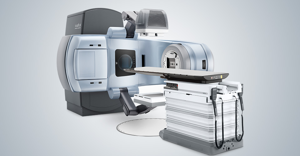
Varian Clinac IX with Rapid Arc
This powerful workhorse treats thousands of patients around the world every day. Designed to deliver a wide range of imaging and patient treatment options, the Clinac system offers advanced features to facilitate state-of-the-art treatments including IMRT, IGRT, RapidArc and stereotactic body radiotherapy.
Rapid Arc delivers the precise dose distribution and conformity of IMRT and IGRT in a fraction of the time, often two minutes or less. By simultaneously shortening treatment times and improving treatment accuracy.
These latest radiation therapy treatment techniques offers advantages of
- Delivering higher dose to the target- tumor thereby increasing cancer control rates
- Decreasing side effects and long term complications by reducing dose to the nearby normal organs such as spinal cord, heart, lungs etc..
HDR Brachytherapy
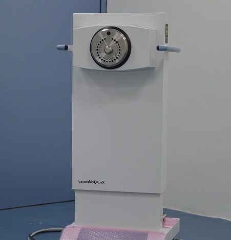
Gammamed plus iX
- 24 channel or 3 channel system
- Unique design ultra-flexible solid core source cable, Iridium-192 source
- High dose rate (HDR) and so shorter treatment time
- OPD treatment, no need to stay in bed for long time
- Intra-cavitary brachytherapy for cancers of uterus and cervix
- Painless and comfortable procedure
- Interstitial implants for soft tissue sarcomas, tongue cancers
Gamma Camera – SPECT
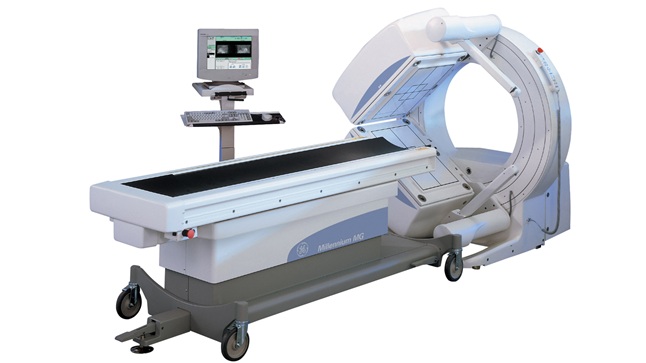
GE-Millenium MG
Millennium MG, Multi-Geometry Nuclear Medicine System is used to perform a range of Nuclear Imaging studies. Imaging with Gamma Camera involves preparation and injection of radiopharmaceuticals that specifically trace the function of Organ of Interest. Radiopharmaceuticals are prepared in the well-equipped Hot Lab in the Department of Molecular Imaging. These specific radiopharmaceuticals are administered to patients and images are acquired. Static, dynamic and parametric images can be obtained under a Gamma Camera.
Diagnostic Services:
- PETCT for initial staging, Detection of Unknown Primary, Radiation therapy Planning, Response assessment, Surveillance
- Sentinel Lymph Node Lymphoscintigraphy
- Radio-guided Occult Lesion Localization (ROLL)
- MDP Bone scan
- MIBG, HYNICTOC and DMSA (V) Imaging for endocrine tumors
- Parathyroid Imaging
- Whole body Iodine Scan for thyroid cancer


